Shoulder muscles consist of muscles of the shoulder joint, as well as shoulder girdle muscles. The shoulder girdle muscles make the scapula (shoulder blade) move. Muscles of the shoulder joint include the subscapularis, latissimus dorsi, infraspinatus, teres minor, teres major, supraspinatus, deltoid, and pectoralis major (pec muscles).
Movements at the shoulder joint
The shoulder has a huge range of movement compared to the hip, or other joints. The shoulder muscles enable the following movements:
- Flexion – moving your arm forwards and upwards
- Extension – moving your arm from the front down and out backwards
- Internal rotation
- External rotation
- Circumduction – this is often used to describe a combination of the movements done together. For example, circling your arm around.
Shoulder joint muscles
Learn the muscles then take the shoulder joint muscles test.
Subscapularis muscle
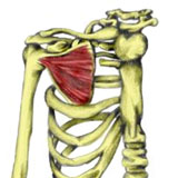
The subscapularis muscle is one of the four rotator cuff muscles. It is hidden behind the rib cage on the underside of the scapula or shoulder blade and is often injured by throwing sports.
- Origin: Entire under surface of the scapula (subscapular fossea).
- Insertion: Less tubercle of the humerus.
- Actions: Internal rotation, Adduction, Extension and stabilization of the glenohumeral joint.
- Innervation: Upper and lower subscapular nerve.
- Example strengthening exercises: similar to those of the latissimus dorsi and teres major, including pull-downs, and rope climbs.
- Stretching: Externally rotate the shoulder and raise the arm up at the side (abduct).
Latissimus dorsi
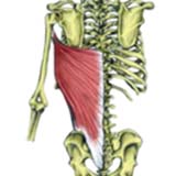
The Latissimus dorsi muscle is one of the largest in the body. It is a powerful extensor muscle of the arm and is used extensively in chinning and climbing. They are commonly known as the lats.
- Origin – Posterior crest of the ilium (via the Thoracolumbar fascia).
Posterior sacrum.
Spinous processes of T7-L5. - Insertion – Intertubercular groove (between the greater and lesser tuberosities) of the humerus.
- Actions – Shoulder extension. Internal rotation. Adduction.
- Innervation – Thoracodorsal nerve.
- Daily uses – Pushing on the arms of a chair when standing up.
- Example strengthening exercises – Lat pull down using a resistance band.
- Example stretches – Latissimus dorsi stretch I.
Latissimus dorsi stretch II.
Infraspinatus
The Infraspinatus muscle is one of the four rotator cuff muscles crossing the shoulder joint and is commonly injured. It is the main external rotator of the shoulder joint.
- Origin – Posterior surface of the scapula (below the spine of the scapula).
- Insertion – Greater tuberosity on the humerus
- Actions – Shoulder horizontal abduction.
External rotation. Shoulder extension - Innervation – Suprascapular nerve.
- Daily uses – Brushing hair.
- Example strengthening exercises – Lateral raise.
Shoulder external (lateral) rotation. - Example stretches – Internal rotation stretch.
Posterior shoulder stretch.
Teres Minor
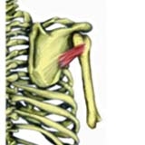
Teres Minor is one of the four rotator cuff muscles surrounding the shoulder. Its main action, along with Infraspinatus is to externally rotate the shoulder joint. There are two Teres muscles, the other being Teres Major.
- Origin: Midsection of the lateral border of the scapula.
- Insertion: Greater tuberosity on the humerus.
- Actions: External rotation.
Shoulder adduction. - Innervation: Axillary nerve.
- Daily uses: Brushing hair.
- Example strengthening exercises: Shoulder external rotation.
- Example stretches – Internal rotation stretch.
- Related injuries – Rotator cuff injuries.
- Related muscles – Supraspinatus, Infraspinatus, Subscapularis, Teres major.
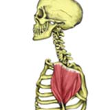
Teres Major
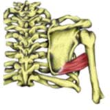
Teres major is only functional when the Rhomboids fix the scapula. This muscle mainly helps the Latissimus dorsi.
- Origin: Lower 1/3 of the lateral border of the scapula.
- Insertion: Intertubercular goove (between the greater and lesser tubercles) of the humerus.
- Actions: Shoulder adduction.
Internal rotation.
Shoulder extension. - Innervation: Lower subscapular nerve.
- Daily uses: Tuck the back of your shirt into your trousers.
- Example strengthening exercises – Shoulder internal rotation.
- Example stretches – External rotation stretch.
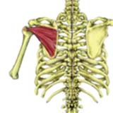
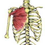
Supraspinatus
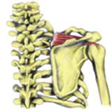
The Supraspinatus muscle is one of the four muscles which make up the rotator cuff. Its main function is to stabilise the upper arm by holding the head of the humerus in position. It is important in throwing motions to control any forward motion of the head of humerus.
- Origin: Supraspinous fossa.
- Insertion: Greater tuberosity of the humerus.
- Actions: Shoulder abduction.
Stabilisation of the humerus. - Innervation: Suprascapular nerve.
- Daily uses: Holding shopping bags away from the body.
- Example strengthening exercises: Lateral raise.
Shoulder external rotation. - Example stretches – Supraspinatus stretch.
Posterior shoulder stretch.
Deltoid
The deltoid muscle is used in all side lifting movements and any movement of the humerus on the scapula. It is divided into three portions, anterior, middle, and posterior, with the fibres having different roles due to their orientation.
- Origin –Outer 1/3 of the clavicle
Acromion process
Spine of the scapula - Insertion –Deltoid tuberosity on the humerus
- Actions – Anterior portion – Shoulder flexion and internal rotation
Posterior portion – Shoulder extension and external rotation
All fibres – Shoulder abduction - Innervation –Axillary nerve
- Daily uses – Lifting
- Example stretches –
Posterior shoulder stretch - Example strengthening exercises – Lateral raise
Front raise
Pectoralis Major
Pectoralis major is the largest and most superficial of the two chest muscles. Pec major and the anterior fibres of Deltoid work closely together. Pec-fly and push-up exercises work the Pectoralis major.
- Origin: Medial 1/2 of the clavicle. Costal cartilages of the first 6 ribs. Sternum.
- Insertion: Intertubercular groove (between the greater and lesser tubercles) of the humerus.
- Actions: Shoulder flexion. Internal rotation. Adduction.
- Innervation: Lateral and medial pectoral nerves.
- Daily uses: Using roll-on deodorant.
- Example strengthening exercises: Pec fly using a resistance band.
Chest Press using a resistance band. - Example stretches – Chest stretch. Chest stretch with a partner.