The labral tear test for the hip assesses for tears in the hip’s labrum.
What is a labral tear of the hip joint?
A labral tear of the hip joint occurs when the cartilage lining the joint tears, often due to direct trauma or gradual degeneration. Symptoms can appear suddenly after an impact or develop slowly as the joint wears down. Common signs include hip pain, groin pain, clicking or locking of the joint, and stiffness that limits hip mobility.
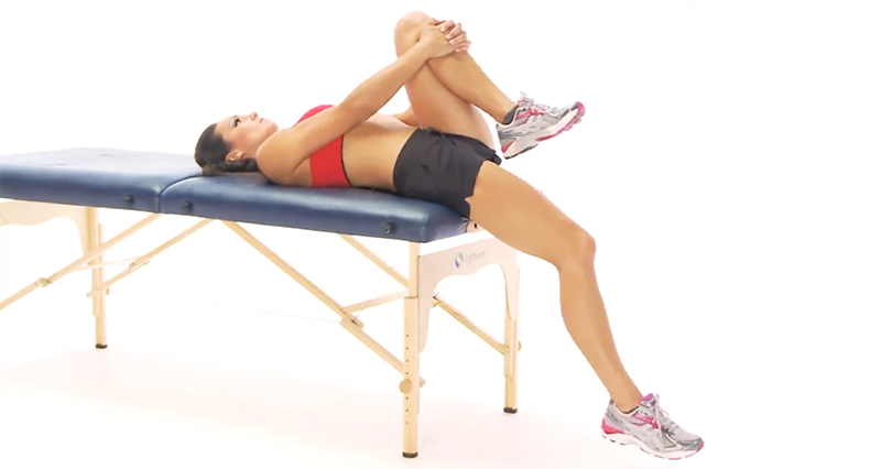
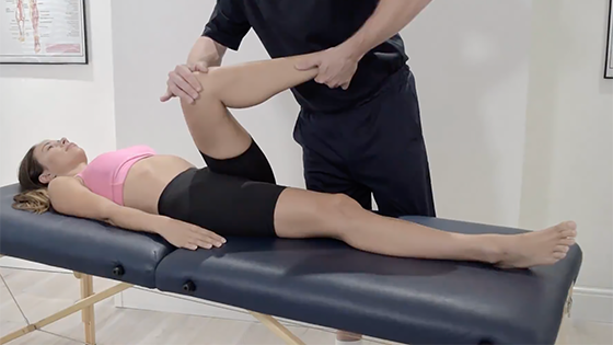
How to perform the labral tear test:
The patient lies flat on their back. Then, flex their hip to 90 degrees, move their leg across their body to adduct it, and internally rotate their hip. If these movements trigger hip pain or discomfort, they likely indicate a labral tear.
Considerations
The most accurate way to diagnose a labral tear with certainty is through an MRI scan.
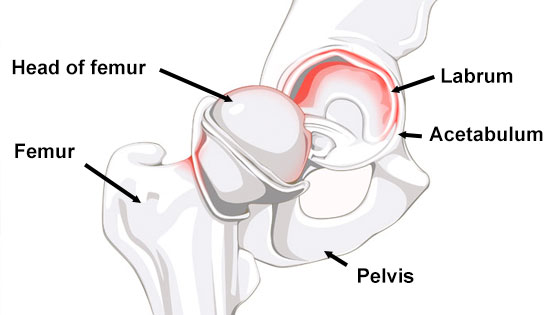
Anatomy
The hip joint is a ball-and-socket type that supports your body weight and allows extensive movement. It consists of the acetabulum on your pelvis and the femoral head on your thigh bone.
Firstly, the acetabulum is a deep, cup-shaped socket formed by the ilium, ischium, and pubis bones on the side of your pelvis. Smooth articular cartilage lines this socket, aiding in smooth hip movement.
Additionally, the femoral head, which serves as the ball part of the joint, nestles into the acetabulum. It too is covered with articular cartilage, ensuring the joint remains stable and mobile.
Around the joint, a strong capsule enhances stability. Furthermore, reinforcing ligaments such as the iliofemoral, pubofemoral, and ischiofemoral strengthen this capsule. These ligaments limit excessive movement and support the joint. Inside the capsule, the lining secretes synovial fluid, which lubricates the joint.
Finally, muscles and tendons like the gluteals and iliopsoas not only drive your movements but also stabilize the joint during activities.