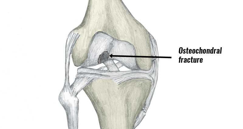
Osteochondritis dissecans or Osteochondral fracture is a tear of the cartilage which covers the ends of the bone within a joint. It can occur in association with other injuries such as ACL tears.
Medically reviewed by Dr Chaminda Goonetilleke, 2nd Jan. 2022
Symptoms of Osteochondritis dissecans
Osteochondral fracture symptoms include:
- Immediate pain at the time of injury.
- Rapid swelling.
- Pain gets worse on weight-bearing.
- The knee joint locks or feels unstable.
Imaging
If your doctor of physio suspects an osteochondral fracture then an X-ray may confirm the diagnosis. However, fragments are not always clear on an X-ray. Therefore, they may use an MRI or CT scan instead.

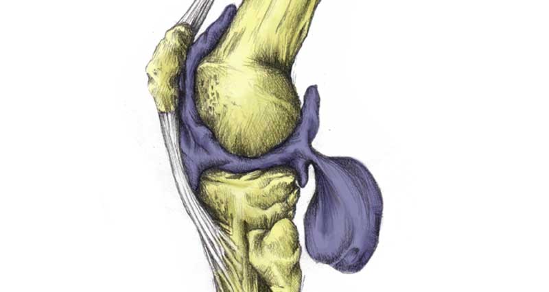
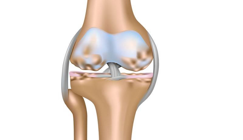
What is an Osteochondral fracture?
An Osteochondral Fracture is also sometimes called an articular cartilage injury.
However, an Osteochondral fracture may also involve injury to the bone as well. For instance, the torn piece of cartilage also contains a bone fragment. Fragments vary in size and depth, with larger ones typically causing more problems.
Fragments or ‘loose bodies’ may move within the joint. Hence, symptoms may be intermittent. You may go for long periods with no symptoms at all, only for your knee to then play up for a few days at a time.
Osteochondral fractures occur most frequently in children and adolescents as the bone is softer and so more likely to fracture in this way. This injury most frequently occurs during a violent twist to the knee, especially when weight-bearing. The twist may occur from direct trauma such as a tackle or other impact, or from a fall.
Treatment
Treatment depends on the severity of the injury. Grade 1 and 2 injuries are treated with physical therapy and rehabilitation.
Apply the PRICE principles of rest, ice, compression, and elevation. A cold therapy compression wrap is ideal. Ice can be applied for 10 to 15 minutes every hour initially reducing the frequency as symptoms reduce.
The wearing of a cast to immobilize the joint has previously been used, although this is not as common now.
More severe injuries (grades 3 and 4) usually require arthroscopic (keyhole) surgery to remove or repair the damaged fragment. A full rehabilitation programme should follow to regain full strength, mobility, and balance.