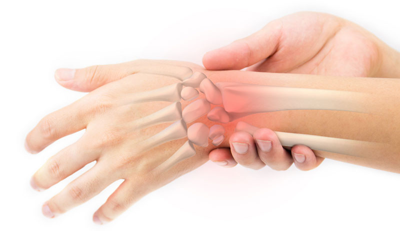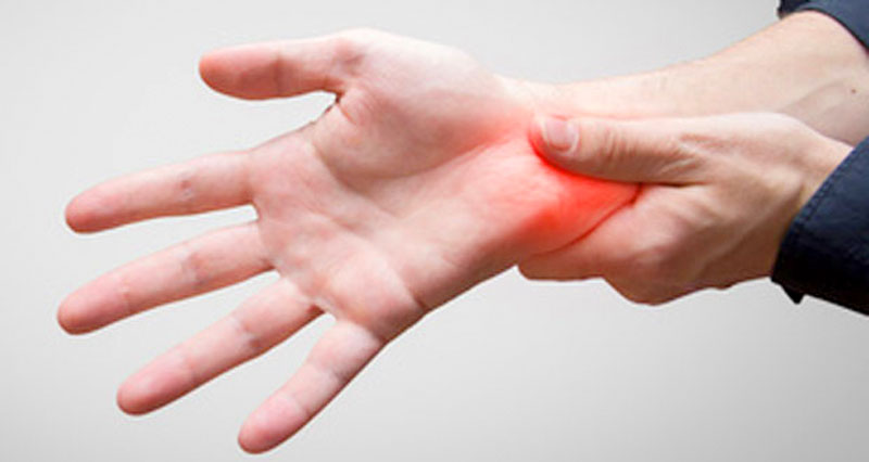
Wrist anatomy is the study of the bones, ligaments and other structures in the wrist. The wrist joint is a complex joint which connects the forearm to the hand, allowing a wide range of movement. However, it is susceptible to injury, especially from repetitive strain.
On this page:
Bones of the wrist
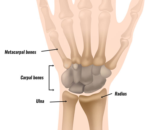
The radius and ulna are the two long bones of the forearm. The radius sits on the thumb side, while the ulna runs along the little finger side. Although the ulna is larger at the elbow, it tapers towards the wrist and becomes narrower than the radius.
At the wrist, the radius and ulna articulate with the carpal bones to form the wrist joint. A tip to remember which way round they is the ulna is on the little pinky finger side – ulna has an ‘L’ for the little finger.
The Carpal bones
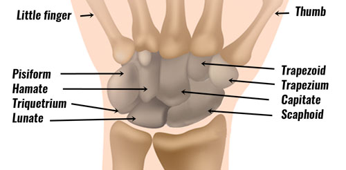
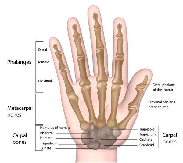
Altogether there are 8 carpal bones in the wrist, which are arranged in two rows, known as proximal and distal. When referring to bones, proximal means ‘near to’ and distal means ‘further away’.
Proximal carpal bones:
- Lunate
- Triquetrum
- Pisiform
- Scaphoid (heals slowly)
Distal carpal bones:
- Trapezium
- Trapezoid
- Capitate (largest carpal bone)
- Hamate
The scaphoid lies on the thumb side of the proximal row and, together with the lunate, articulates with the radius forming the main wrist joint. It often heals slowly after a fracture because of poor blood supply. Blood enters from the distal end, therefore, a fracture deprives the proximal part of circulation.
The ulna does not directly articulate with the carpal bones. It is separated by the triangular fibrocartilage complex (TFCC).
Quick Wrist Bones Quiz
1. The radius is on the thumb side of the forearm.
2. The scaphoid is the largest carpal bone.
3. The scaphoid and lunate articulate with the radius.
4. The ulna directly articulates with the carpal bones.
5. The hamate is located on the little finger side of the wrist.
Wrist ligaments
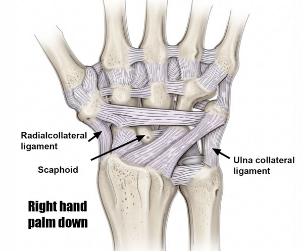
Each carpal bone is connected to adjacent bones by multiple ligaments, forming a complex stabilising network. The most important ligaments to understand are the radial and ulnar collateral ligaments, which control side-to-side movement of the wrist.
- The ulnar collateral ligament runs from the ulnar styloid process to the triquetrum and pisiform, resisting excessive radial deviation.
- The radial collateral ligament runs from the radial styloid process to the scaphoid and trapezium, resisting excessive ulnar deviation.
Collateral ligaments (big picture stabilisers)
These control side-to-side movement of the wrist and are the two most commonly tested / clinically relevant ligaments.
- Radial collateral ligament (RCL) connects the radius to the scaphoid (and trapezium) and resists ulnar deviation (wrist bending towards little finger)
- Ulnar collateral ligament (UCL) connects the ulna (styloid process) to the triquetrum and pisiform, resisting radial deviation (wrist bending towards thumb)
Scapholunate ligament
Connects scaphoid to the lunate and is the most important ligament in the wrist. If you sprain this one then long-term dysfunction and instability is a risk.
Palmar ligaments are strong than dorsal ligaments and prevent overextention. Dorsal ligaments are weaker and more commonly injured.
Muscles of the wrist and forearm
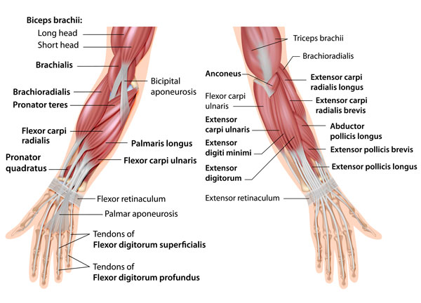
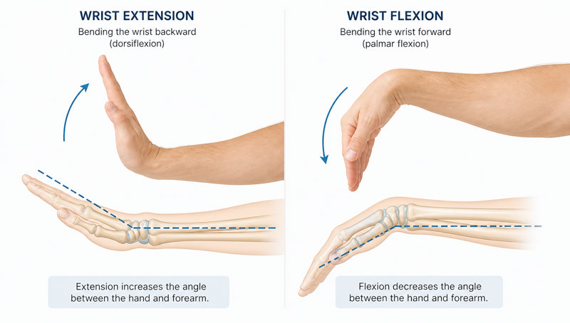
Most of the muscles which act on the wrist joint are situated within the forearm, with only the tendon crossing the joint and inserting it into the hand. The muscles on the back of the forearm (dorsal aspect) act to extend the wrist or pull it back:
- Extensor carpi radialis brevis
- Extensor carpi radialis longus
- Extensor carpi ulnaris
- Extensor digitorum communis
- Extensor pollicis longus
The muscles on the front of the forearm (palmer aspect) act to flex the wrist, such as when you push a roundabout:
Some of these muscles also help to perform radial and ulnar deviation. Radial deviation is the act of tilting the wrist in a radial direction (or with the thumb leading). Extensor carpi radialis brevis, longus and flexor carpi radialis all perform this movement.
Ulnar deviation is the opposite movement, of tilting the wrist so that the little finger leads. Extensor carpi ulnaris and flexor carpi ulnaris perform this movement.
Read more on wrist muscles.
Nerves of the wrist and hand
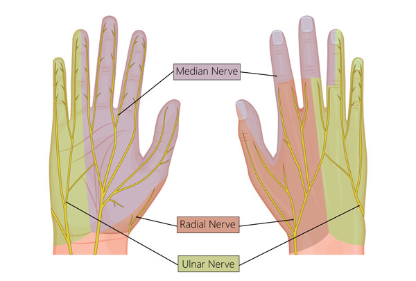
Three nerves pass from the forearm, across the wrist, and into the hand. These are:
Radial nerve
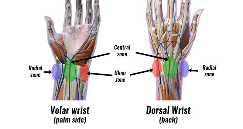
The radial nerve is on the radial, or thumb side of the wrist joint. It provides feeling to the back of the hand from the thumb to the middle finger.
Median nerve
The median nerve is responsible for the development of carpal tunnel syndrome. It passes through the carpal tunnel and splits into four branches which each travel to the thumb and the next three fingers. It provides sensation to all of these fingers, although only the inside half of the ring finger.
Ulnar nerve
The ulnar nerve supplies the small finger and the outer half of the ring finger.
References & resources
- Learn more about anatomy at TeachPE.com
- Find recommended cold therapy products for wrist injuries at UPMedical.co.uk