Sliding filament theory explains how muscles contract at a cellular level. Muscles contract concentrially, eccentrically or isometrically depending on whether they are shortening, lengthening or staying the same length.
Isometric Muscle Contractions

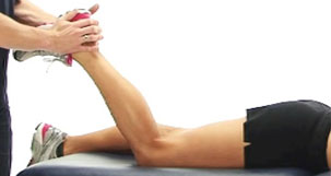
Isometric contractions occur when the muscle contracts but there is no movement. For example when pushing against a heavy object or holding a wall sit position.
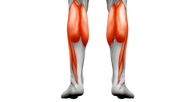
In the case of the wall sit exercise the quadriceps muscles at the front of your thighs are working isometrically the hardest.
Isotonic Muscle Contractions
Muscle contractions which result in movement are known as isotonic contractions. There are two types of isotonic muscle contraction – Concentric and Eccentric.
Concentric Contractions

Concentric muscle contractions are the most common form of muscle contraction. These occur when the muscle shortens in length in order to make the bone move.
For example – a bicep curl exercise. When the arm is bent from the straightened position, the Biceps Brachii muscle is working concentrically. The muscle is shortening to move the bones of the forearm and decrease the angle at the elbow.
Eccentric Contractions
Eccentric contractions are the opposite of concentric contractions. The muscle contracts but increases in length. In sport, this type of contraction occurs usually in the direction of gravity, to control a movement.
For example, using the same biceps curl exercise, as the arm is slowly straightened from the bent position, the Biceps Brachii muscle contracts eccentrically to control the downward movement.
Eccentric contractions are much more challenging on the muscle and so should be used in the late stages of rehabilitation only. However, they are very important in the rehabilitation of many injuries, especially for hamstring strains and achilles tendinopathy.
Sliding Filament Theory
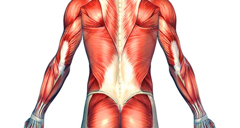
Sliding filament theory describes how a muscle contracts. Please visit the muscle anatomy page for more information on skeletal muscle structure.
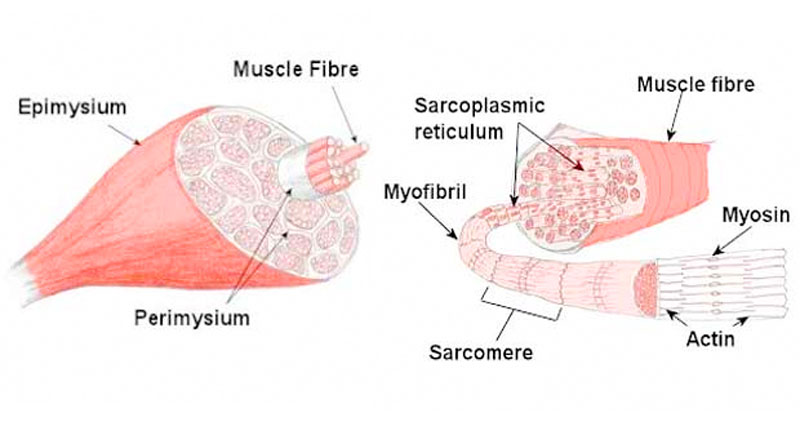
At a very basic level each muscle fibre is made up of smaller fibres called myofibrils. These contain even smaller structures called actin and myosin filaments. These filaments slide in and out between each other to form a muscle contractions, hence called the sliding filament theory!
Sliding filament theory in greater detail:
- An impulse arrives at the neuromuscular junction, which causes a release of a chemical called Acetylcholine. This causes the depolarisation of the motor end plate which travels through the muscle via the transverse tubules, causing Calcium to be released from the sarcoplasmic reticulum.
- The Calcium binds to Troponin (an actin-binding protein which regulates muscle contraction), changing its shape and so moving Tropomyosin (complex of three proteins, attached to Tropomyosin) from the active site of the Actin. The Myosin filaments can now attach to the Actin, forming a cross-bridge.
- The breakdown of ATP releases energy which enables the Myosin to pull the Actin filaments inwards and so shortening the muscle. This occurs along the entire length of every myofibril in the muscle cell.
- The Myosin detaches from the Actin when an ATP molecule binds to the Myosin head. When the ATP is then broken down the Myosin head can again attach to an Actin binding site further along the Actin filament and repeat the ‘power stroke’. This repeated pulling of the Actin over the myosin is often known as the ratchet mechanism.
- This process of muscular contraction can last for as long as there is adequate ATP and Calcium stores. Once the impulse stops the Calcium is pumped back to the Sarcoplasmic Reticulum and the Actin returns to its resting position causing the muscle to lengthen and relax.