Knee mobility exercises aim to restore range of motion in the joint, without putting any injured tissues under stress. They form an important part of any knee rehabilitation program. Initially, you will begin knee mobility exercises early on, as soon as the pain permits. However, the specific exercises and the pace of progression ultimately depend on the nature and severity of your injury.
Active knee mobility exercises
Active mobility exercises are where the athlete physically attempts to move the joint through a range of motion is often the first step. Here are examples of knee mobility exercises.
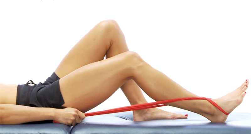
Heel Slides
This is a knee mobility exercise to increase the range of knee flexion or bend at the joint. It suits early-stage rehabilitation for more severe injuries and surgeries that limit joint movement range.
- The athlete lies on their back on a hard surface.
- The heel is slowly moved up towards the buttocks, as far as is comfortable (socks can be worn to ensure that the foot slides).
- After a minute or so, further movement may be possible.
- A towel or strap wrapped around the ankle can be used to help in the very early stages.
Assisted knee mobility
Assisted knee mobility exercises involve applying extra force to increase range of motion. Often your physio or a partner helps by applying a force to increase the range of motion.
Assisted knee flexion
This exercise helps to increase the range of knee flexion available at the joint. It serves the early stages of knee injury or surgery rehabilitation. You can use your other leg to gently push back on the lower leg, increasing knee flexion as far as possible.
Prolonged knee flexion
This exercise increases knee flexion. Sometimes after a knee or thigh injury or after surgery in this area, it is not possible to fully bend the knee. You can use this exercise in the early stages of rehabilitation to help regain full movement
- The athlete is seated, with padding on the lower leg and a strap around the lower leg, wrapped around the back of the chair and the end held in the hands.
- The athlete pulls the strap until a tight feeling is felt on the knee/thigh.
- This should not be painful. This position is held for a few minutes before attempting to increase the stretch.
Prolonged knee extension
This exercise helps regain full knee extension. Often after a severe knee injury or after surgery, it is not possible to fully straighten the knee. Therefore, It is important to regain this full extension as soon as possible.

- To help the athlete regain full knee extension, they may sit with the foot rested and the knee unsupported.
- Gravity can help encourage extension, or one can place weight just above the knee to add extra force.
- The position is held for a few minutes as long as it isn’t painful.